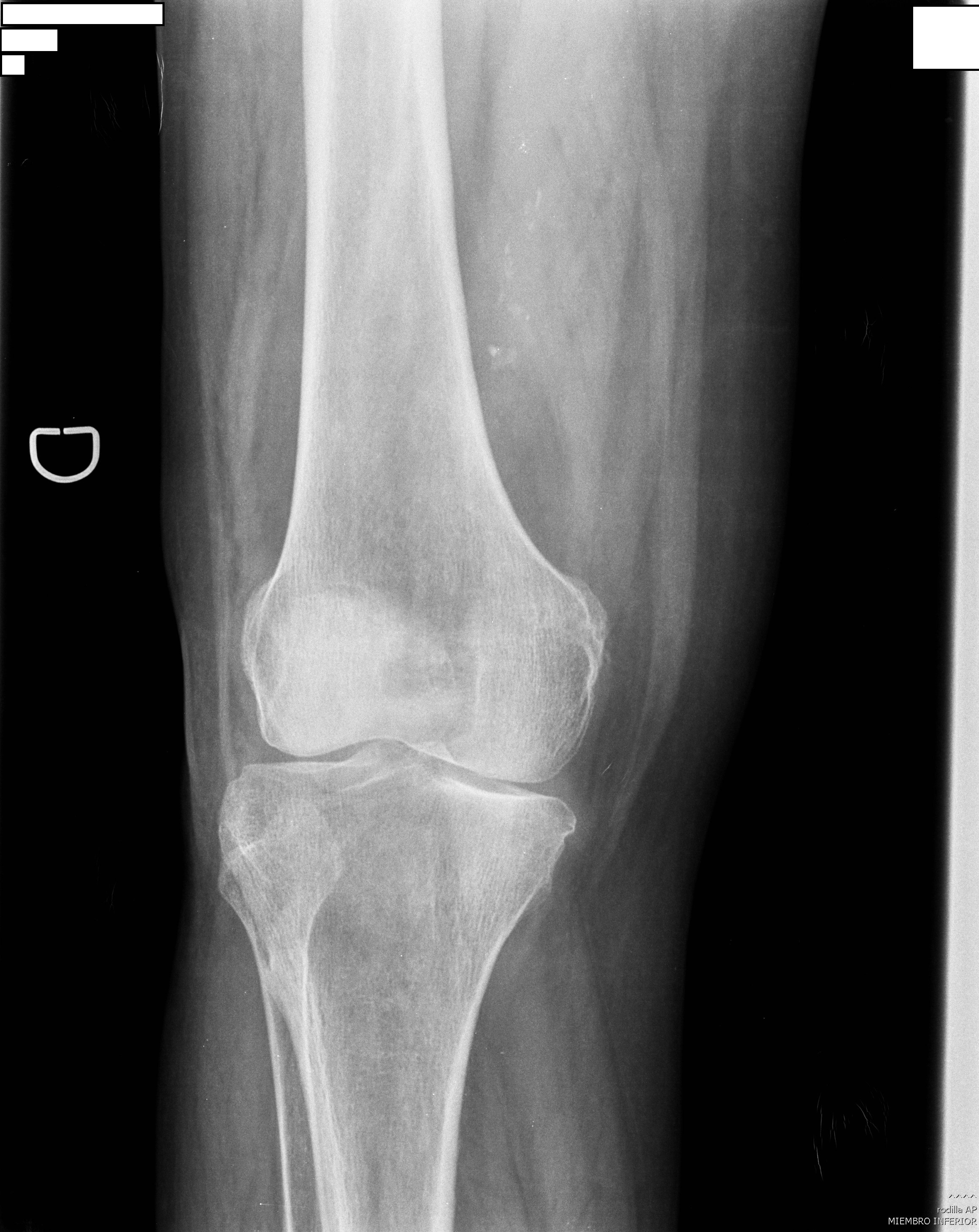
AP KNEE PROJECTION
Anteroposterior • Frontal View • Femorotibial Joint Evaluation
Exposure Factors
Equipment: Without or with bucky. Position: Supine decubitus.
Plate Size
CENTERING POINT
Anatomical reference point for cassette and central ray centering
Visible Anatomical Structures
Distal Femur
Inferior portion of femur
Proximal Tibia
Superior portion of tibia
Proximal Fibula
Superior portion of fibula
Patella
Overlapping in AP view
- Knee joint - Femorotibial joint
- Femoral condyles - External and internal of femur
- Intercondylar tubercle - Medial and lateral
- Internal tibial tuberosity - Ligament insertions
- Joint space - Femorotibial interarticular line
- Tibial plateaus - Tibial articular surfaces
- Intercondylar eminence - Tibial spine
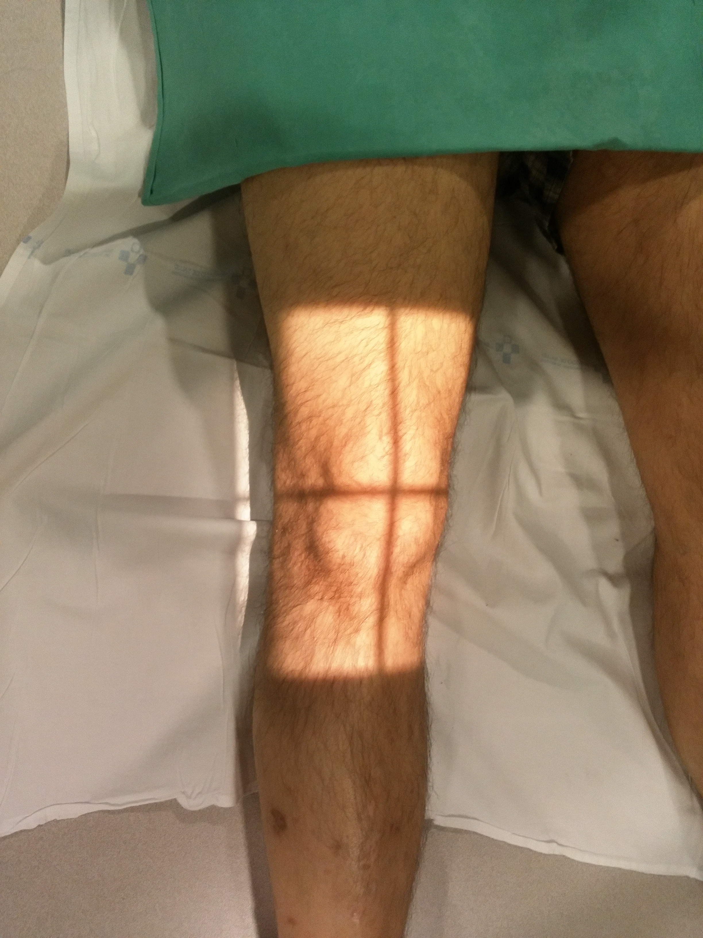
Patient Positioning
Central Ray Direction
Directed to 1 cm below patella apex
Entry point: Anterior, at patella level
Exit point: Posterior, intercondylar space
Cephalic angulation: Optional, for better joint space visualization
Patient Instructions
"Remain still during the examination"
Do not move leg during exposure - Maintain full extension
Technical Considerations
Slight Rotation
Internal rotation of foot to align condyles parallel to cassette.
Cephalic Angulation
5-10° cephalic optional for better joint space visualization.
Bone Inclusion
Include sufficient proximal and distal diaphysis for complete evaluation.
Clinical Indications
Image Quality Criteria
Joint Space
Femorotibial joint space symmetrical and visible
Condyle Symmetry
Femoral condyles aligned and symmetrical
Bone Inclusion
Sufficient proximal and distal diaphysis included
Complete Knee Study
STANDARD RADIOGRAPHIC SERIES
The AP knee projection is typically complemented with: